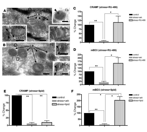
Figure 5. PS-induced decrease in AMP delivery to epidermal LBs is reversed by RU-486.
(A and B) Ultrathin sections labeled with CRAMP (A) and mBD3 (B) primary antibodies followed by a 10-nm colloidal gold–tagged secondary antibody after embedding for electron microscopy. Sections were postfixed in osmium tetroxide and embedded in LR White medium. Black arrows denote unlabeled LBs; circles indicate label in cytosol. (A) CRAMP was labeled (black arrowheads) in nonstressed normal controls (Co), and CRAMP labeling reappeared in PS mice treated with RU-486 (PS+Ru), but not with exogenous lipids (PS+L). (B) Exogenous lipids restored labeling of mBD3 in LB in PS mice (black arrowheads). Scale bars: 100 nm. (C–F) Quantitative data for immunolabeling of CRAMP and mBD3 in LB in nonstressed control or PS mice plus either RU-486 (C and D) or lipid (E and F) cotreatment. *P < 0.05; **P < 0.001.

