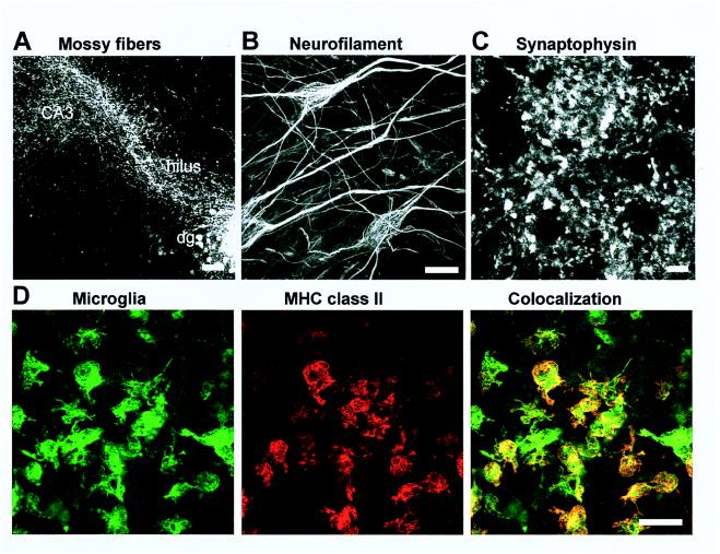
Figure 1.
Neuronal outgrowth and MHC class II expression of microglia in the cultured hippocampal slices analyzed by confocal laser-scanning microscopy. Neuronal outgrowth was determined by anterograde labeling of mossy fibers innervating the hilus and the CA3 region of the hippocampus (A), by immunolabeling for the cytoskeleton protein neurofilament (B), and by immunolabeling for the synapse protein synaptophysin (C). Slices were treated with IFN-γ (50 units/ml) and double-labeled for MHC class II and isolectin for microglia (D). MHC class II molecules were visualized mainly on microglia. [Bars = 100 μm (A), 20 μm (B), 5 μm (C), 50 μm (D).]

