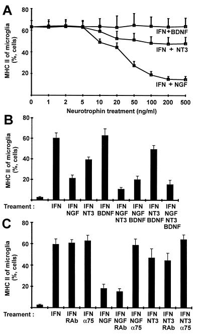
Figure 4.
Analysis of MHC class II expression of isolated cultured microglia by flow cytometry. (A) Isolated microglial cells were treated for 72 hr with IFN-γ (IFN, 50 units/ml) and different concentrations of NGF, BDNF, and NT3 for 72 hr. (B) Isolated microglial cells were induced by IFN-γ (IFN, 50 units/ml), and different combinations of NGF (50 ng/ml), BDNF (50 ng/ml), and NT3 (50 ng/ml) were added to the cell cultures for 72 hr. (C) In addition, microglial cells were treated for 72 hr with IFN-γ (IFN, 50 units/ml), and action of NGF (50 ng/ml) and NT3 (50 ng/ml) on MHC class II of microglia was inhibited with rabbit antibody blocking the p75 neurotrophin receptor (α75). Isolated microglial cells were treated with rabbit antibody (RAb) as a control. Data are presented as relative number of cells (mean and SEM).

