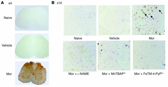
Figure 2. Micrographs of the superficial layers of the dorsal horn of the lumbar enlargement (L4–L6) of the spinal cord illustrating nitrotyrosine staining.
(A) On day 5 acute injection of morphine (3 mg/kg) or its vehicle did not lead to the appearance of nitrotyrosine staining in the superficial layers of the dorsal horn. On the other hand, acute administration of morphine on day 5 after repeated administration of morphine led to significant protein nitration in the superficial layers of the dorsal horn with no staining in the ventral horn. Original magnification, ×4. (B) Coadministration of morphine over 4 days with l-NAME (10 mg/kg/d), MnTBAP3– (10 mg/kg/d), or FeTM-4-PyP5+ (30 mg/kg/d) blocked nitrotyrosine formation. Original magnification, ×10. Micrographs are representative of at least 3 from different animals in experiments performed on different days.

