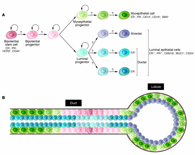
Figure 3. Hypothetical model of human mammary epithelial stem cell hierarchy and differentiation.
(A) Hypothetical depiction of mammary epithelial stem cells and their various progeny. A bipotential stem cell gives rise to luminal epithelial and myoepithelial cells, but the intermediary steps and their regulation are largely unknown (question marks). The model is likely to oversimplify the real situation, since there are many different types of luminal epithelial cells and both the myoepithelial and luminal cells are likely different in the ducts and alveoli. (B) Schematic picture of a normal terminal duct lobular unit with the putative location of the various stem and differentiated cells indicated. Gray line denotes the basement membrane; color of cell types correlates with that in A. CK14, cytokeratin 14; MUC1, mucin 1.

