
Fig. 1. The MBT domains of dSfmbt, hSFMBT and hL(3)MBT are conserved.
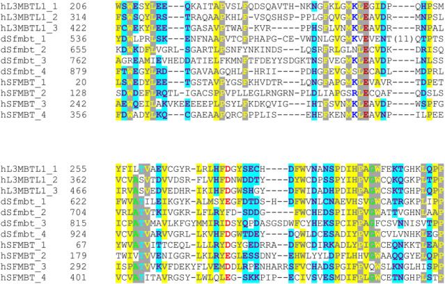
Sequences comprising the MBT repeats of each protein were obtained from the SMART Domain Database [40]. Sequences were aligned using ClustalX [41] and rendered using CHROMA [42]. Residues sharing sequence identity within the MBT repeats are denoted with a gray background, while conserved hydrophobic and hydrophilic residues are illustrated with yellow and cyan backgrounds, respectively. Conserved acid residues are red. The MBT repeats of fly and human SFMBT are 34% identical and 48% homologous.