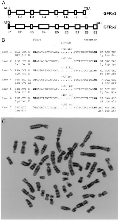
Figure 2.
Genomic analysis of GFRα3. (A) Intron–exon junctions in the coding region of GFRα2 and GFRα3. Exons are represented by boxes and scaled according to size. Introns (intervening lines) are not to scale. Note that GFRα3 entirely lacks one exon relative to GFRα2. (B) Precise intron sites in the mouse GFRα3 gene. Consensus splice sites are shown in boldface type. (C) Fluorescence in situ hybridization analysis of the human GFRα3 gene location. Digital image of a metaphase chromosome spread derived from methotrexate-synchronized normal human peripheral leukocytes after hybridization with a digoxigenin-labeled GFRα3 genomic probe and 4′,6-diamidino-2-phenylindole counterstaining. Both copies of chromosome 5 have symmetrical rhodamine signals on sister chromatids at region 5q 31.2–32.

