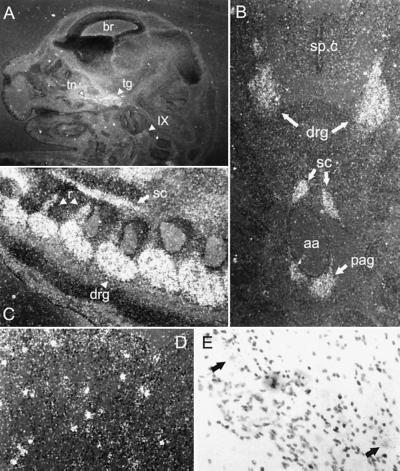
Figure 4.
In situ hybridization analysis of GFRα3 expression in mouse. (A) Saggital section of E14 mouse head. GFRα3 is observed in trigeminal ganglion (tg) and nerve (tn) and in the glossopharyngeal ganglion (IX) but not in brain (br). (B) Transverse section of E14 mouse showing GFRα3 expression in DRG but not in the spinal cord (sp.c). Around the abdominal aorta (aa), staining is observed in the sympathetic chain ganglia (sc) and the preaortic ganglia of Zuckerkandl (pag). (C) Saggital section of E14 mouse spinal column showing DRGs (drg), sympathetic chain (sc), and labeled nerve roots (r). (D) Dark-field photomicrograph of adult trigeminal ganglion showing punctate staining in contrast to the diffuse staining observed at E14. (E) Bright-field photomicrograph of adult trigeminal ganglion at higher power, showing GFRα3 is localized to neurons (arrows).

