Figure 5.
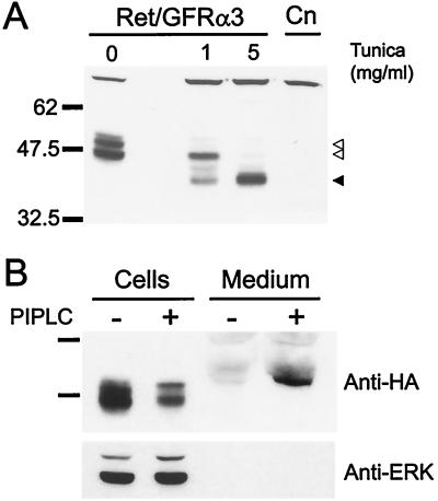
Analysis of NHA-GFRα3 protein in transfected fibroblasts. (A) Anti-HA immunoblot of fibroblasts stably transfected with HA-tagged GFRα3 and Ret (Ret/GFRα3) show a doublet at approximately 47 and 51 kDa (open arrowheads) that was not present in the parental line (Cn). Tunicamycin (Tunica) treatment of the cells for 24 hr to block N-linked glycosylation resulted in loss of the upper band at 1 mg/ml, and appearance of a lower molecular mass band. At a tunicamycin concentration of 5 mg/ml, only the lower band running at the predicted molecular mass for GFRα3 was visible (solid arrowhead). (B) Phosphatidylinositol-specific phospholipase C (PIPLC) treatment of NHA-GFRα3 expressing fibroblasts to specifically cleave GPI-linked proteins resulted in depletion of the NHA-GFRα3 from the cells and induced the presence of a band in the medium corresponding to NHA-GFRα3. Reprobing of the blot with an anti-ERK p42/44 antibody is shown below to indicate equal loading of cell lysates.

