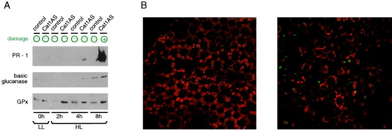
Figure 5.
Necrosis-independent expression of defense proteins in Cat1AS plants. (A) Western blot analysis with PR-1, bPR-2, and GPx antibodies. Cat1AS and control plants were exposed to HL for various times (2–8 h) and then returned to LL for 2 weeks before leaf sampling. Expression of PR-1, bPR-2, and GPx was induced by 4 h of HL in Cat1AS, whereas necrosis required 8 h of exposure. PR-1 expression in the control line was not increased by HL. bPR-2 and GPx showed HL induction in the control line but not to the same level as in Cat1AS. Leaf damage was assessed at the time of harvest: no damage indicates that none of the leaves of that plant had any visible sign of injury. (B) Optical sections of a YOYO-1 iodide-stained leaf from a Cat1AS plant treated with HL for 4 h (Left) or 8 h (Right). YOYO-1 iodide is a nuclear dye that is membrane-impermeant and therefore stains only nuclei of damaged cells. (×700.)

