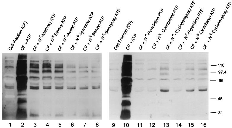
Figure 2.
Anti-phosphotyrosine protein immunoblot showing the level of protein tyrosine phosphorylation after treatment of a murine lymphocyte cell lysate (CF) with 100 μM of ATP or A*TPs (analogs 1–12). This lysate includes Src, Fyn, Lck, Lyn, Yes, Fgr, Hck, Zap, Syk, Btk, Blk, and other tyrosine kinases present in B and T lymphocytes, macrophages, and follicular dendritic cells (28). Molecular size standards (in kilodaltons) are indicated.

