Figure 3.
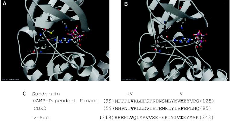
(A) A close-up view of the ATP binding site in cAMP-dependent protein kinase (1ATP). Three residues within a 4-Å sphere of the N6 amine of ATP (Val 104, Met 120, and Glu 121) in 1ATP and the catalytically essential lysine residue (Lys 72) are shown in ball-and-stick representation. The remainder of the protein is shown in ribbon format. (B) cdk2–cyclin D complex (1cdk) cocrystal structure. The 1cdk structure is shown with bound ATP, the residues in the 5-Å sphere of the N6 amine of ATP (Val 64, Phe 80, and Glu 81) and the catalytic lysine residue (Lys 33) are shown in ball-and-stick representation. A and B were created by feeding the output of molscript into the raster3d rendering program (35, 36). (C) Sequence alignment of the ATP binding regions of PKA, cdk2, and v-Src. The bold residues correspond to the amino acids with side chains in a 5-Å sphere of the N6 amino group of kinase-bound ATP.

