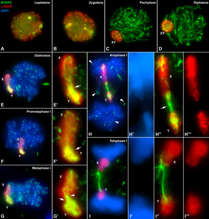
Figure 5. Immunolabeling of Squashed Spermatocytes with Anti-SYCP3 (Green) and Anti γ-H2AX (Red) Antibodies and Counterstaining of Chromatin with DAPI (Blue).
Several focal planes have been projected in each image.
(A) Leptotene: γ-H2AX labeling appears distributed throughout the whole nucleus.
(B) During zygotene γ-H2AX distribution is very similar to that observed in the previous stage.
(C) From pachytene onwards, the bulk of anti-γ-H2AX antibody is located onto the sex chromosomes, which appear located at the nuclear periphery and remain so during diplotene (D).
(E–E′) Diakinesis: Sex chromosomes appear distally connected and intensely labeled with γ-H2AX. Note the SYCP3 connection between both arms of the sex chromosomes (arrow in E′). The same situation is found in prometaphase I (F–F′).
(G–G′) Metaphase I: The autosomal bivalents remain aligned at the metaphase I plate. The connection between the sex chromosomes has broken in the X chromosome short arm (arrowhead), and only the long arm is linked to the Y (arrow).
(H–H′′′) Anaphase I: Sex chromosomes migrate to the poles, but they are delayed compared to autosomes. Note the remnants of SYCP3 over the autosomes and dispersed in the cytoplasm (arrowheads). A thick SYCP3 filament is visible between the X and Y chromosomes (arrow), but no chromatin joining is detected as revealed by DAPI (H′) or γ-H2AX staining (H′′′).
(I–I′′′) Telophase I: Sex chromosomes are clearly lagged in segregation. The SYCP3 filament appears connecting them without chromatin connection (I′–I′′′).

