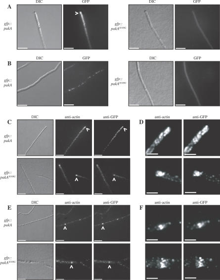
Figure 1. PakA Co-Localises with Actin at Sites of Polarised Growth.
Localisation of PakA in live cells (A, B) and in immunofluorescently labeled fixed cells (C–F) at 25 °C. (A) The GFP::PakA fusion protein is localised to the hyphal apex at 25 °C (white arrowhead). In contrast, the GFP::PakAH108G fusion protein is visible throughout the cytoplasm. (B) In subapical hyphal cells, the GFP::PakA fusion protein is observed as spots along subapical hyphal cells. This localisation is not observed in the gfp::pakAH108G strains. (C) The PakA and PakAH108G GFP fusion proteins co-localised with actin at the hyphal tip (indicated by white arrowheads). (D) Enlargement of the stained region indicated by arrowheads in C. (E) At 25 °C the PakA and PakAH108G GFP fusion proteins also co-localise with actin at nascent septation sites (indicated by arrowheads). (F) Enlargement of the stained region indicated by arrowheads in E. Images were captured using DIC or under epifluorescence to detect GFP, anti-actin, or anti-GFP. Scale bars, 20 μm.

