
Figure 5.
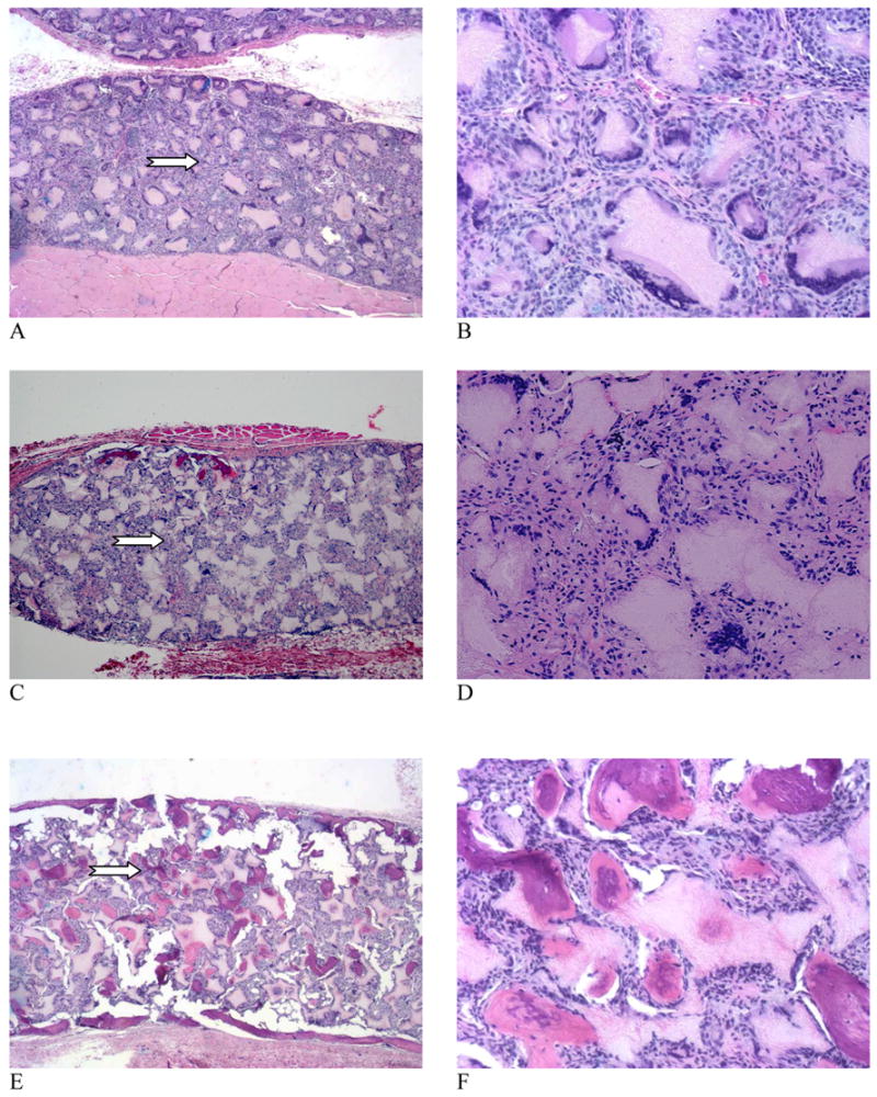
Microscopic observations of the H & E stained tissue sections of scaffolds retrieved 6 weeks after implantation. (A, B) Control scaffold; (C, D) 5 μg rhBMP-7 adsorbed to scaffold; (E, F) 5 μg rhBMP-7 incorporated in NS-scaffold. Original magnifications: (A, C, E) 40x full cross sections, and (B, D, F) 200x for high magnification views of selected representative areas (arrows point to the selected areas in A, C, and E). Note: In order to conduct von Kossa staining (Figure 6), the engineered tissue samples were not decalcified and the sectioning resulted in some artifacts, which appeared in the H&E histology (multiple cracks in the highly mineralized constructs, i.e., E & F).