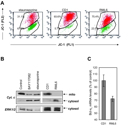
Fig. 2.
PrPSc-induced mitochondrial disruption leads to apoptosis. A. JC-1 staining of staurosporine, CD1 (control) and RML6 cells. Cells treated with staurosporine (1 μM, 4 h), CD1 or RML6 for 18 h were stained with JC-1 according to the protocol and analysed on a BD FACS Calibur™ as described in Experimental procedures. JC-1 fluorescence is seen in both the FL-2 (red) and FL-1 channels (green). JC-1 that fluoresces in the FL-1 channel and lacks fluorescence in the FL2 channel is considered to correspond to mitochondria with a depolarized ΔΨ. For staurosporine-treated cells 67% showed decreased fluorescence in the FL-2, while only a small proportion of the population in CD1 control cells (5.7%) showed depolarization of the ΔΨ. After RML treatment 23.5% of the cells mitochondria were found with a depolarized ΔΨ. B. Western blot analysis of mitochondrial (mito) and cytosol fraction of untreated and either staurosporine-, BAY117082-, CD1- or RML6-stimulated Bos2 cells, using an antibody directed against cytochrome c. Equal loading was controlled by detection of ERK1/2 in the cytosol fraction. C. Real time qPCR to detect murine bcl-xl from RML6- versus CD1-treated cells. The expression of bcl-xl normalized to 18S RNA and the SEM for triplicate experiments are given in percentage of the control (CD1). Statistic analysis using the Mann–Whitney rank sum test showed significant differences between RML6- and CD1-treated cells (P = 0.028).

