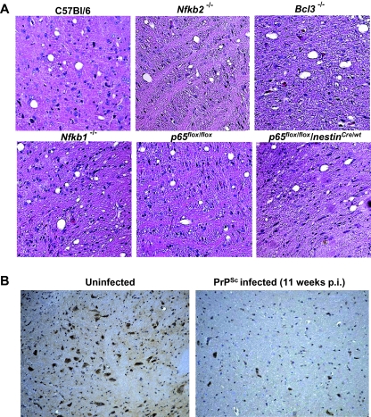
Fig. 6.
Lesions in the brain and p65 detection of PrPSc-infected mice. A. Terminally sick mice showed comparable vacuolation (haematoxylin/eosin staining, H&E) after i.c. inoculation of PrPSc. The pons areas of midsagittal sections are shown. Magnification ×20. Three mice of each strain were analysed. B. Strong reduction of activated NF-κB (phospho-p65) in PrPSc-infected C57Bl/6 mice. Uninfected and infected animals at 11 weeks p.i. were sacrificed and the brain of three animals per group were analysed by immunohistological staining using a phospho-p65-specific antibody. Representative pons areas of midsagittal sections are shown. Magnification ×10.

