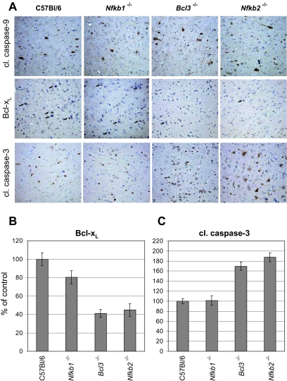
Fig. 7.
Reduced Bcl-xL expression correlates with enhanced caspase-3 activation in NF-κB2- and Bcl-3-deficient mice after 11 weeks of PrPSc infection. A. Bcl-xL expression (black arrows), active cleaved caspase-9 and caspase-3 were determined by immunohistological staining. Bcl3–/–, Nfkb2–/–, Nfkb1–/– and C57Bl/6 mice were inoculated with RML6 and three animals per groups were sacrificed at 11 weeks p.i. Brain sections of three animals were analysed for each mouse line. Representative areas of the entorhinal cortex are shown. Magnification ×20. B and C. For quantification of positive cells, three sections of three different mouse brains for each strain were chosen. The graphic represents the amount of positive cells in the brain of the deficient mice compared with the C57Bl/6 mice in percentage of control. SEM was shown as bars. For detailed information on quantification see Experimental procedures.

