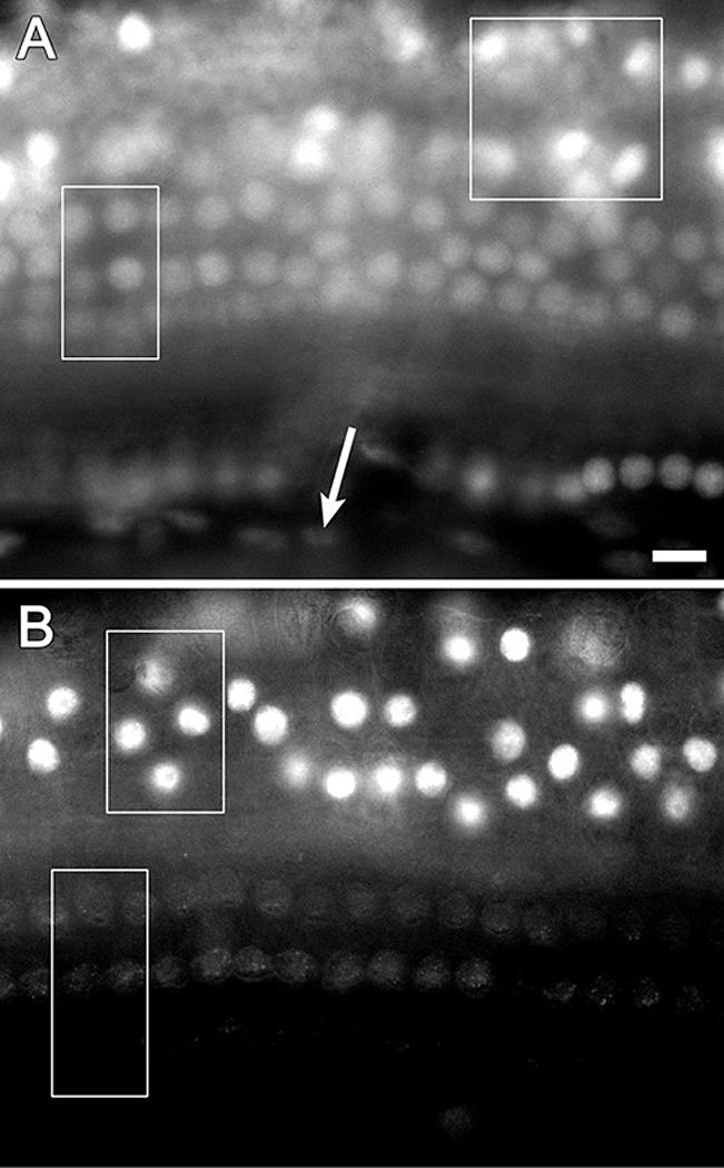
Figure 1.

The normal distribution of p27Kip1 in mature guinea pig ears shown by immunofluorescence on whole-mounts of the auditory epithelium. A. At a focal plane immediately above the basilar membrane positive nuclei are found in Deiters cells (rectangle), Hensen cells (square) and inner pillar cells (round nuclei at bottom of image). Spindle shaped mesothelial cells located beneath the basilar membrane display background level staining (arrow). B. At a higher focal plane, p27Kip1 positive nuclei are found in Hensen cells (top rectangle) whereas nuclei of hair cells are at background staining level (bottom rectangle). Bar, 10 μm for A and B.
