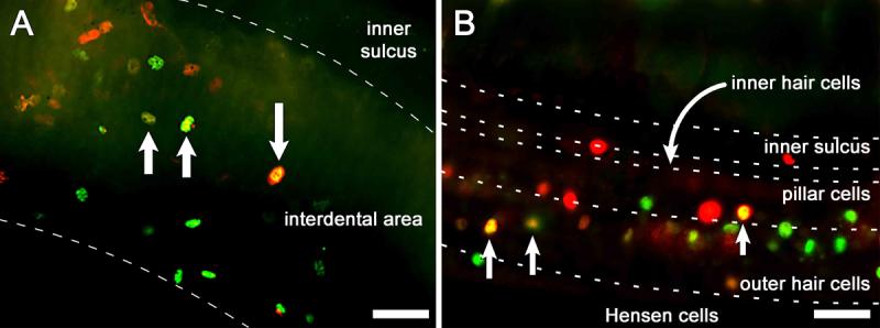
Figure 3.

Epi-fluorescence images of whole-mounts of the organ of Corti double immuno-labeled for Atoh1 (green) and SKP2 (red) 4 days after Ad.Atoh1 and Ad.SKP2 inoculation. Images were obtained immediately beneath the luminal surface. A. In the interdental cell region, numerous cells are stained for Atoh1 or SKP2, and a small number of cells (yellow) express both proteins (arrows). B. In the organ of Corti and Hensen cell area, several cells (arrow) are yellow indicating dual expression of SKP2 and Atoh1 while others express either Atoh1 (green) or SKP2 (red). Dashed lines delimit regions within the tissue. Bar, 20 μm.
