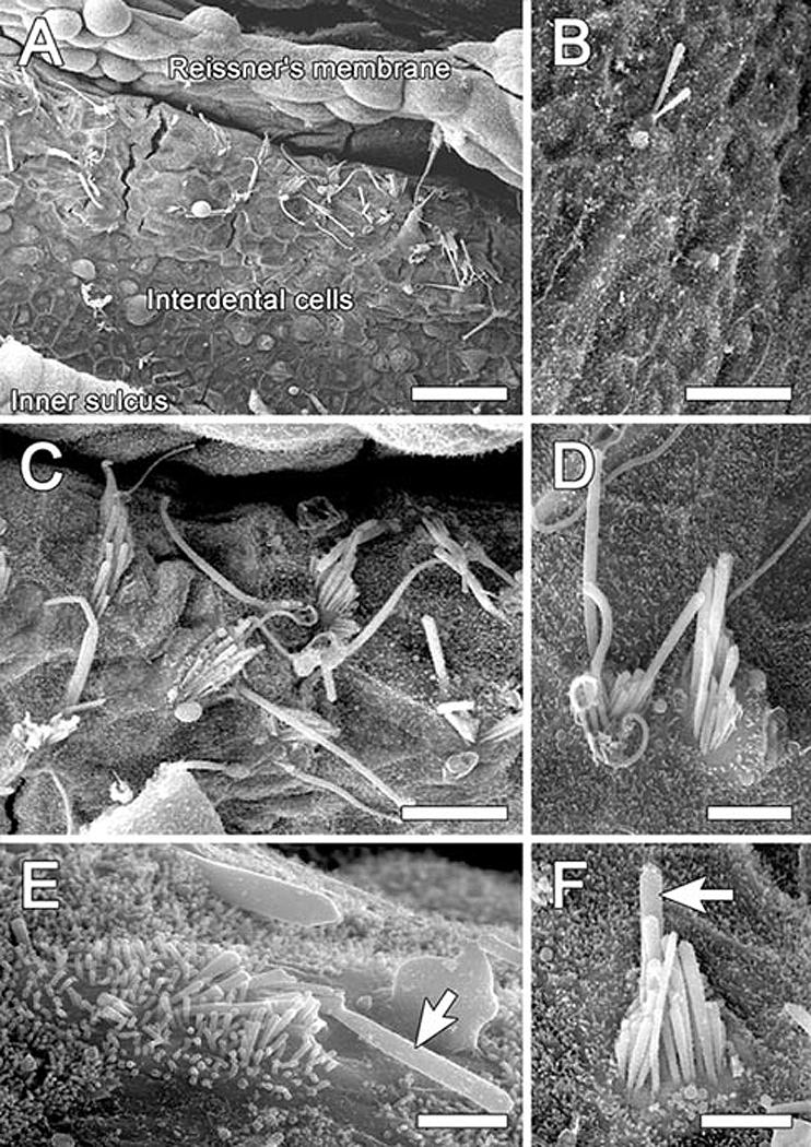
Figure 4.

SEM images of interdental cell area (2nd turn) 2 months after inoculation of Ad.Atoh1 and Ad.SKP2 (A and C-F) or Ad.Atoh1 alone (B). A. Numerous ectopic stereocilia bundles on the limbus, reaching to the area where Reissner's membrane is inserted. B. A single ectopic bundle among interdental cells on the limbus. C. An enlarged area in (A) showing stereocilia bundles in an ectopic location on the limbus. Some bundles contain a graded array of stereocilia and others are rather disorganized. A kinocilium-like projection is seen on some of the bundles. D. Two ectopic hair cells appearing as a pair . E. Some ectopic hair cells appear like immature cells and exhibit a long and/or thick kinocilium-like protrusion (arrow). F. An ectopic hair cell with a staircase organization of stereocilia and a projection that appears like a kinocilium (arrow). Bars, 30 μm in A, 10μm in B, C, 5μm in D, 2 μm in E, and 3 μm in F.
