
Figure 4.
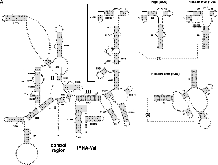
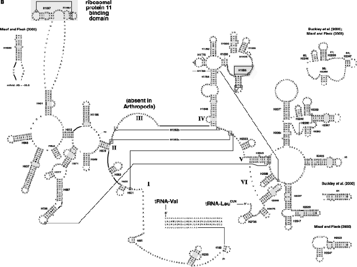
The secondary structure model of the mitochondrial rRNA (12S + 16S) from the honey bee, Apis mellifera. Differences between our sequence and previously published A. mellifera sequences (U65190 and U65191) are in bold, with insertions (dark arrows), deletions (open arrows) and substitutions (parentheses) shown. Helices aligned across all sampled panarthropods are boxed in grey. Tertiary interactions (where there is strong comparative support) and base triples are shown connected by continuous lines. (A) SSU rRNA (12S). Misaligned sequences 1 and 2 (discussed in text) are within dashed boxes and connected to redrawn structures from Hickson et al. (1996) and Page (2000) with dashed lines. (B) LSU rRNA (16S). See Fig. 2 legend for explanations of base pair symbols, helix numbering and reference for software used to construct structure diagrams.