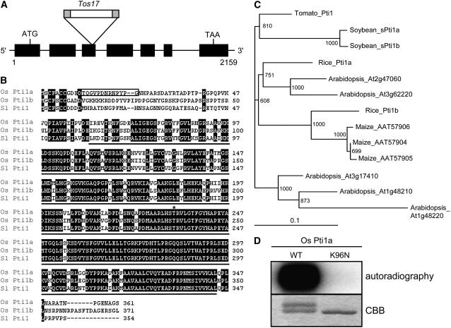
Figure 3.
Comparison of Os Pti1a and Related Proteins.
(A) Relative position of the Tos17 insertion within the Os Pti1a gene. Exons are indicated by the black boxes.
(B) Alignment of the predicted amino acid sequences of Os Pti1a, Os Pti1b, and Sl Pti1. A black line below Sl Pti1 indicates a conserved protein kinase domain, and the box delimits the peptide sequence used to derive Os Pti1a–specific antisera. The asterisk marks the T233 phosphorylation site of Sl Pti1 by Pto and the corresponding sites in Os Pti1a and Pti1b.
(C) A phylogenetic tree constructed with the amino acid sequences of kinase domain of Os Pti1a and Pti1 family members from several plant species.
(D) Autophosphorylation assay for Os Pti1a (WT) and its mutant form (K96N) in vitro. The upper band in the wild-type lane seen in the Coomassie blue (CBB)–stained gel is presumably the phosphorylated form.

