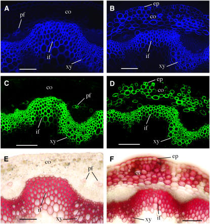
Figure 10.
Deposition of Cellulose, Xylan, and Lignin in the Ectopic Secondary Walls in MYB46 Overexpressors.
The stem sections were stained with Calcofluor White or phloroglucinol-HCl for detection of cellulose and lignin, respectively. The LM10 xylan monoclonal antibody was used for immunodetection of xylan. co, cortex; ep, epidermis; if, interfascicular fiber; pf, phloem fiber; xy, xylem. Bars = 105 μm.
(A) and (B) Calcofluor White staining of stem sections showing intensive cellulose staining in the walls of cortical cells and epidermis in addition to interfascicular fibers and xylem cells in MYB46 overexpressors (B) compared with the wild type (A).
(C) and (D) Stem sections probed with the LM10 xylan monoclonal antibody showing intensive xylan staining in the walls of cortical cells and epidermis in addition to interfascicular fibers and xylem cells in MYB46 overexpressors (D) compared with the wild type (C).
(E) and (F) Phloroglucinol-HCl staining of stem sections showing intensive lignin staining in the walls of cortical cells and epidermis in addition to interfascicular fibers and xylem cell walls in MYB46 overexpressors (F) compared with the wild type (E).

