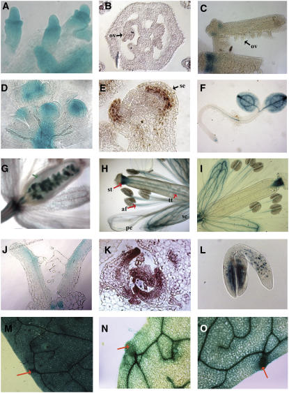
Figure 2.
Expression Analysis of SAW and BEL1 Genes.
(A) Immature ovules showing BEL1:GUS activity.
(B) SAW1 in situ hybridization of a cross section through the gynoecium showing immature ovules (ov).
(C) SAW2:GUS expression in whole-mount gynoecia. One of the gynoecia was split open to reveal the immature ovules.
(D) Whole mount of the inflorescence apex. BEL1:GUS expression is detected in the floral meristems of immature flowers.
(E) Longitudinal section of a stage 4 flower showing SAW1 expression (assayed by in situ hybridization) in the adaxial side of the developing sepal (se).
(F) SAW2:GUS expression in a 2-d-old seedling.
(G) to (I) Whole mounts of flowers showing BEL1:GUS, SAW1:GUS, and SAW2:GUS activity, respectively. st, style; tt, transmitting tract; af, anther filament; se, sepal; pe, petal.
(J) to (L) BEL1, SAW1, and SAW2 exhibit adaxial expression in developing lateral organs.
(J) A longitudinal section through the vegetative apex of a 7-d-old seedling showing adaxial expression of BEL1:GUS in the leaves.
(K) Cross section through the vegetative apex showing in situ localization of SAW1 in the adaxial side of developing leaves.
(L) SAW2:GUS expression in a developing embryo.
(M) to (O) Whole mounts of fifth leaves of 3-week-old plants showing BEL1:GUS, SAW1:GUS, and SAW2:GUS activity, respectively.

