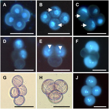
Figure 3.
Pollen development analysis of T1 AGL18∷AGL18 RNAi plants forming 50% nonviable pollen in a qrt1-2 background. Representative microspores, isolated from anthers at different uninucleate and binucleate stages and stained with 4′,6-diamidine-2′-phenylindole dihydrochloride (1 μg/mL dissolved in PBS), are shown. A, Early uninucleate microspore stage. Due to the qrt1-2 background, microspores still stick together, revealing that wild type-like nuclei are located in the center of the cells. B and C, At the late uninucleate microspore stage, formation of big vacuoles (arrows) in two microspores moves nuclei from the center to the cell wall. The two other microspores are lacking big vacuoles and likely degenerated later. D, Nuclei disappeared in two microspores; others just completed the first mitotic division. E, At the two-cell stage, two microspores proceeded further in development and generative cells are visible (arrowheads). F, Only wild type-like microspores underwent second mitosis and contain two sperm cells and one big vegetative cell; the two other pollen grains were aborted. G and H, Bright-field images of Figure 3, D and F (respectively). J, Four wild type-like pollen grains in the quartet at a mature stage. Bar = 25 μm. [See online article for color version of this figure.]

