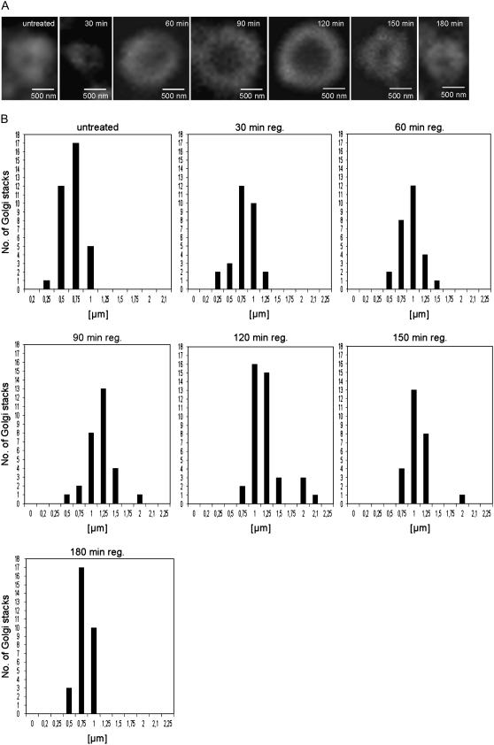
Figure 1.
Golgi stack regeneration during BFA washout in BY-2 cells as monitored by immunofluorescence labeling with ARF1 antibodies. A, Representative individual images from the recovery time points indicated. B, The external diameters of stacks visualized end-on were determined from at least 50 stacks from different cells in samples removed from cultures at the recovery times indicated.

