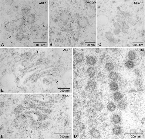
Figure 3.
Immunogold electron microscopy with COPI and COPII antisera. Sections of BY-2 cells prepared by high-pressure freezing/freeze substitution and stained with ARF1, γ-COP for the presence of COPI proteins, and SEC13 for COPII proteins. Samples were removed and freeze-fixed after 20 min (A–D) and 60 min (E and F). c = cis; t = trans.

