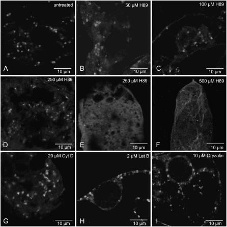
Figure 6.
Effects of inhibitors on Golgi stack regeneration. BY-2 cells were treated with BFA (10 μg mL−1) for 45 min, then allowed to regenerate Golgi stacks in the presence or absence of the indicated inhibitor for a 120-min period. Golgi stack presence was monitored by ARF1 immunofluorescence. Images presented are from 45-μm optical sections in the confocal laser scanning microscope. A, Control cells. B to F, Increasing concentrations of H-89. G and H, Microfilament inhibitors cytochalasin D (20 μm) and latrunculin B (2 μm). I, Microtubule inhibitor oryzalin (10 μm).

