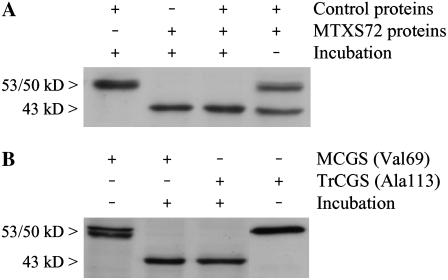
Figure 6.
Evidence for posttranslational cleavage of the N-terminal region of MCGS in folate-deficient cells. A, Soluble proteins prepared from control cells were mixed with an equal amount of proteins from cells treated with MTXS for 72 h (MTXS72) and incubated for 2 h at 25°C. Forty micrograms of proteins were analyzed in each lane. B, Pure recombinant CGS enzymes (25 ng) were incubated for 2 h at 25°C with 2.5 μg soluble proteins from MTXS72 cells. Two versions of the enzyme were analyzed: the MCGS (starting with Val-69) and a truncated CGS (TrCGS, starting with Ala-113) bearing a deletion of 44 residues at the N terminus of the protein. Each combination from sections A and B was analyzed by western blot with polyclonal antibodies raised against CGS. Note that in B only the recombinant CGSs are detected because they are present in excess as compared to the enzyme provided by the MTXS72 extract.

