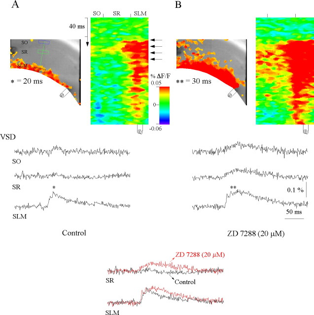
Figure 4.
Intrinsic Ih properties spatially restrict propagation of temporoammonic EPSPs. A, Control. A snapshot of activation at 20 ms of the voltage-sensitive dye (VSD) responses of evoked EPSPs (asterisk) to a burst stimulus (4 stimuli at 100 HZ) in stratum lacunosum moleculare (left) and the activation profile (right) generated from the raster line scan along the path of interest (green line) (see Materials and Methods). VSD SO, SR, and SLM are the local voltage-sensitive dye signals quantified from regions of interest in stratum oriens (blue box), stratum radiatum (green box), and stratum lacunosum moleculare (black box), respectively. The voltage-sensitive dye signals were averaged over 12 trials. Note that the temporoammonic-evoked EPSP is spatially restricted to the extreme distal dendrites of CA1 pyramidal neurons. B, Perfusion of the Ih antagonist ZD 7288 (20 μm) results in significant facilitation of temporoammonic EPSPs to stratum radiatum and stratum oriens (**p ≤ 0.05, ANOVA; n = 4). Left, Snapshot at 40 ms. The inset compares the voltage-sensitive dye responses in stratum lacunosum moleculare and stratum radiatum in control (black) and in the presence of ZD 7288 (red).

