
Figure 5.
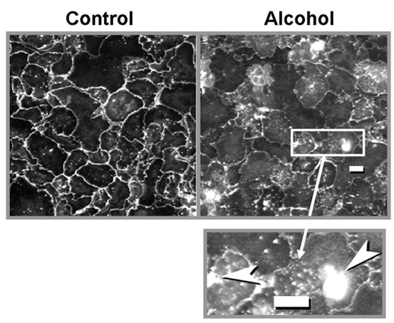
Immunocytochemical localization of claudin-7 protein (see text for details), in alveolar epithelial monolayers from control-fed rats (left panel) and alcohol-fed rats (right panel). As is evident in these representative images, claudin-7 protein is localized primarily to the cell membranes in the control monolayers on the left. In contrast, alcohol monolayers showed patchy localization with many areas where little or no claudin-7 could be visualized in cell membranes. The inset in the lower right is a magnified image of part of the right panel, and illustrates the areas of intracellular accumulation of claudin-7 that were seen in the monolayers from alcohol-fed rats. The size bar in the right panel and in the inset represents 10 microns.