
Fig. 5.
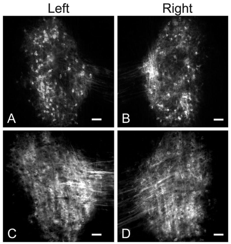
(A, B) Paired images of retrogradely labeled neurons in the left (A) and right (B) DNLL of a control animal after DiI placement in the dorsal tegmental commissure (of Probst) (see experimental design in Fig. 1). Note the symmetric labeling on the left and right side. Scale bars=100 μm. (C, D) Paired images illustrating retrogradely labeled neurons in the contralateral (C) and ipsilateral (D) DNLL after DiI placement in the dorsal tegmental commissure (of Probst) of an ablated animal (see experimental design in Fig. 1). Note the labeling is symmetric on each side although it is more difficult to resolve individual cells than in A and B. Scale bars=50 μm.