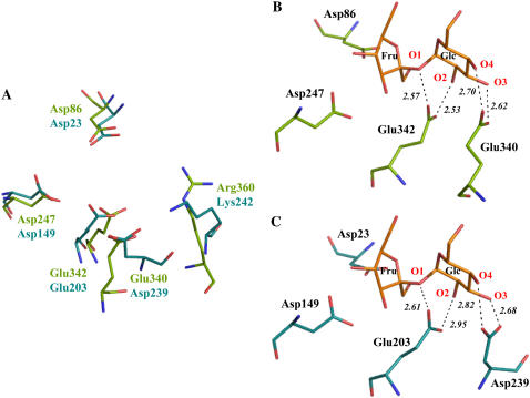
Figure 6.
A, Superposition of the active sites of AtcwINV1 (turquoise) and B. subtilis levansucrase (green; PDB code 10YG) showing similarities in the catalytic region. B, Details of Suc (orange) in the active site of B. subtilis levansucrase Glu-342A mutant (PDB code 1PT2) in which Ala-342 was replaced in silico by Glu-342 (wild-type orientation; PDB code 1OYG). C, Illustration of the putative position of Suc in the active site of AtcwINV1 based on the known structural complex of B. subtilis levansucrase with Suc. Putative Suc-protein interactions are indicated with dashed lines. Figures were prepared with PyMol (Delano, 2002).

