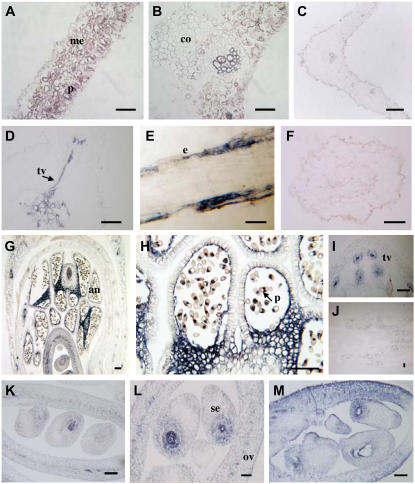
Figure 7.
Localization of TRX m in pea plants by in situ hybridization with the TRX m antisense probe. Bars = 100 μm. A and B, Cross and longitudinal sections of a leaf. C, Leaf section hybridized with the sense probe. D, Cross section of a root. E, Longitudinal section of a root. F, Sense control of TRX m mRNA in roots. G, Longitudinal section of a flower. H, Pollen grain. I, Vascular tissue of the pedicel. J, Longitudinal section of a flower showing the sense control. K to M, Higher magnifications of longitudinal sections of ovaries showing details of developing embryos. me, Mesophyll; p, parenchyma; co, collenchyma; vt, vascular tissue; e, epidermis; al, anther locus; po, pollen; ov, ovary; es, embryonic sac.

