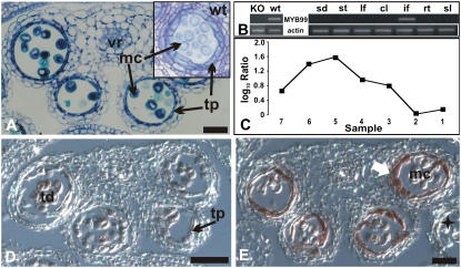
Figure 7.
Characterization of a T-DNA insertion line for MYB99. A, Transverse section through a myb99 mutant anther. Cells are morphologically similar to their counterparts in wild-type anthers (see inset), with the exception of tapetal cells at the vacuolated microspore stage. A representative image is shown of multiple sections of mutant and wild-type anthers that were analyzed. B, MYB99 expression in distinct organs or tissues (sd, Seedling; st, mature stamen; lf, leaf; cl, cauline leaf; if, inflorescence; rt, roots; sl, siliques) of wild-type plants (right) and in whole inflorescences of the myb99 mutant line (KO) compared to the wild type (wt; left). Top, Gene-specific primers for MYB99 were used for RT-PCR; bottom, primers for actin were used as a control to verify that a similar amount of cDNA was used in the different reactions. MYB99 transcripts were only detected in inflorescences. C, Microarray data for MYB99. Log10-transformed ratios from the analysis of temporal gene expression in ms1 mutant flowers are shown. MYB99 expression peaks at early stages of stamen development. D and E, Results of in situ hybridizations for MYB99. D, Transverse section through a stage 7 anther. Only weak hybridization signals were obtained. E, Transverse section of an anther at stage 9 showing expression of MYB99 exclusively in the tapetum (arrow). A star indicates a lateral anther at a later stage of flower development, in which MYB99 expression was not detectable in the tapetum. mc, Microspores; pg, pollen grain; tp, tapetum; td, tetrads; vr, vascular region. Scale bars = 30 μm.

