
Figure 2.
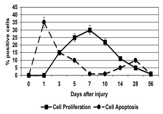
The time course of cell proliferation as determined by BrdU staining and cell apoptosis as determined by TUNEL in wire injured femoral vessels. Values are the mean± s.e.m (n=6 per group). Statistical differences between groups were tested with a Kruskal-Wallis non-parametric test with post hoc Dunn’s multiple comparison correction, where appropriate.