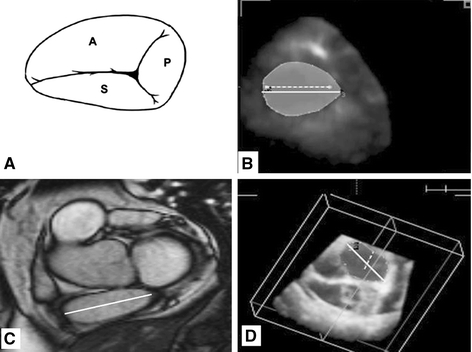
Fig. 1.

Tricuspid annulus oval shape as seen by graphic representation (A), real-time three-dimensional echocardiography (B), magnetic resonance imaging (C), and tricuspid annulus with 2 lines inside, the larger is the TAD by real-time three-dimensional echocardiography and the smaller is the TAD by two-dimensional echocardiography with large difference in measurements
