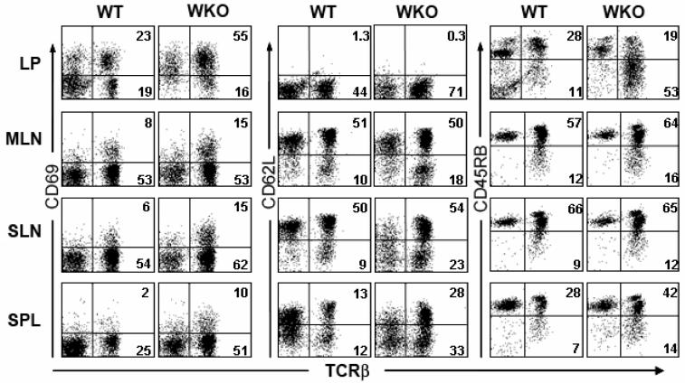
Figure 3. WKO colonic LP contains activated T cells.
Lymphocytes from WT and WKO colonic lamina propria (LP), mesenteric lymph nodes (MLN), subcutaneous lymph nodes (SLN), and spleen (SPL) were analyzed for activation markers. WKO LP is comprised of a higher percentage of T cells (i.e., TCRβ+) than WT. There is marked expansion of activated T cells (as evidenced by CD69+ and CD62L− staining) in the LP and less striking expansion in other lymphoid compartments in WKO mice compared to WT mice. Data are representative examples of at least three individual experiments. Mice used in the experiment were 3-4 months old.

