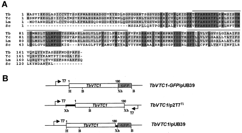
Figure 1. Sequence alignment of TbVTC1.
(A) ClustalW sequence alignment of TbVTC1 (Tb, AAX70699) with S. cerevisiae Phm4/Vtc1 (Sc, P40046) and two orthologues from T. cruzi (Tc, AY304574) and L. major (Lm, CAB 86965). Lines above the sequence represent predicted transmembrane domains. Identical residues are shaded. (B) Scheme of constructs used for GFP localization (upper), RNAi (middle) and overexpression (bottom) experiments. The arrows show the T7 promoters; the ◆ shows the introduced stop codon between TbVTC1 and GFP and the open boxes show the ORFs of TbVTC1 and GFP respectively. The bold line represents the 5′-untranslated region of TbVTC1. B, BamHI; H, HindIII; Xb, XbaI; Xh, XhoI.

