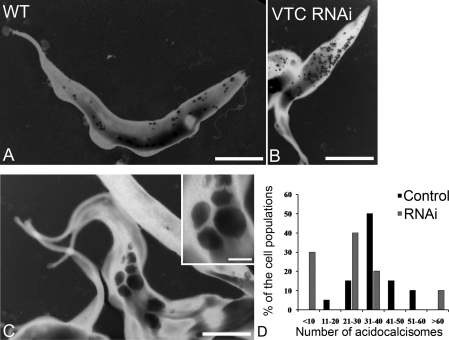
Figure 5. Morphological changes after RNAi of TbVTC1 and numeric distribution of acidocalcisomes in T. brucei.
(A–C) TEM of whole procyclic trypomastigotes. The dark granules are the acidocalcisomes. (A) Wild-type (WT) procyclic stages; (B and C) procyclic forms after 4 days of induction with tetracycline. (B) shows cells with numerous small acidocalcisomes, whereas (C) and inset show cells with large acidocalcisomes. Scale bars: (A) 3 μm, (B) 5 μm and (C) 4 μm. (D) Whole unfixed parasites were observed using a Zeiss EM 902 transmission electron microscope equipped with an energy filter and the number of acidocalcisomes per cell in ∼50 cells was counted.

