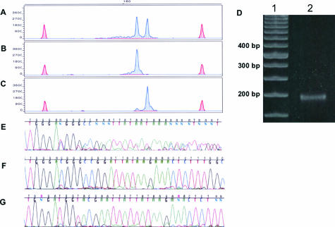
Figure 5.
Isolation of DNA fragments differing in size by three bases. A: TCR PCR products subjected to capillary electrophoresis with an initial voltage of 15,000 demonstrated a clonal pattern of TCR gene rearrangement presenting with two dominant peaks at 183 and 186 bases and a minute amount of polyclonal background. The reconfigured gel block was installed at 22 minutes, voltage was decreased to 5000 V, and collection tubes were changed at 15-second intervals. Reamplification of the isolated DNA fragments revealed a dominant 183-base peak from tube 10 (B) and a dominant 186-base peak from tube 12 (C). TCR peaks are blue-shaded, and size standard peaks are red-shaded. x axis, size in bases. y axis, RFUs. D: Polyacrylamide gel (10%) electrophoresis. Lane 1, 50-bp DNA ladder. Lane 2, TCR gene rearrangement containing two dominant peaks at 183 and 186 bases by capillary electrophoresis that comigrate as a single band on PAGE. E: Sequencing of the pre-isolated PCR product showing mixed and hence unreadable data as expected in the PCR amplicons. F and G: Sequencing of the reamplified purified fractions documenting that they have been sorted to sufficient purity to obtain a single readable sequence.

