
Figure 5.
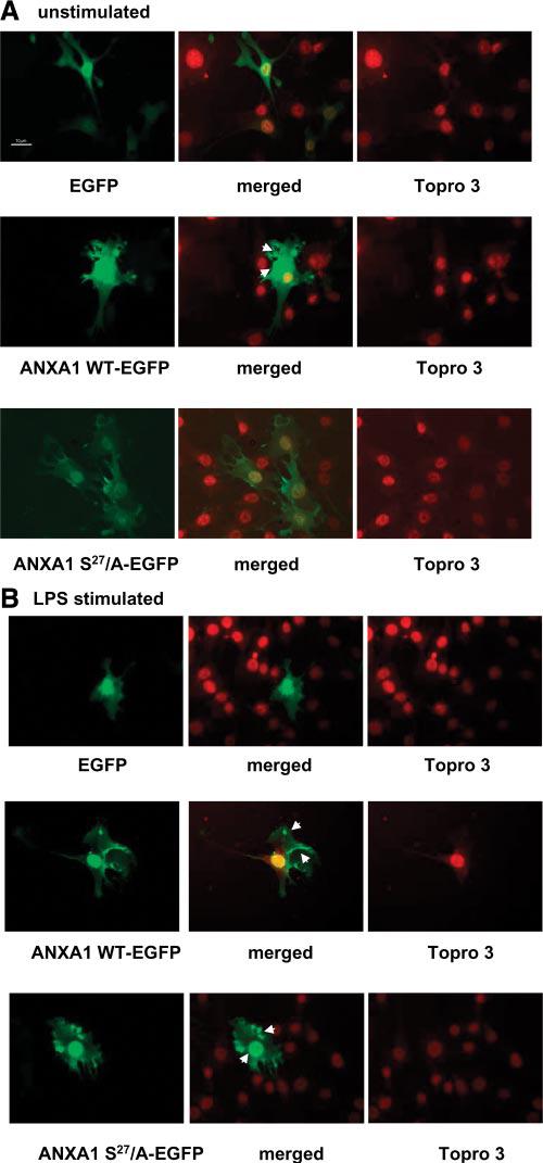
Cellular localization of ANXA1-wild-type-enhanced GFP and ANXA1-S27/A-enhanced GFP mutant in TtT/GF cells. A) Unstimulated cells transfected with empty plasmid EGFP, ANXA1 WT-enhanced GFP, or ANXA1-S27/A-enhanced GFP. Nuclei were stained with topro 3. Merged images represent the EGFP-topro3. Cells were fixed and moviol mounted. B) Cells stimulated with LPS (100 ng/ml, 5 min). All images were collected at similar exposure time. All image files were processed for presentation using Image Pro plus software (Media Cybernetics) and imported directly in PowerPoint in publication format. Arrows indicate area of ANXA1 localization after LPS treatment.