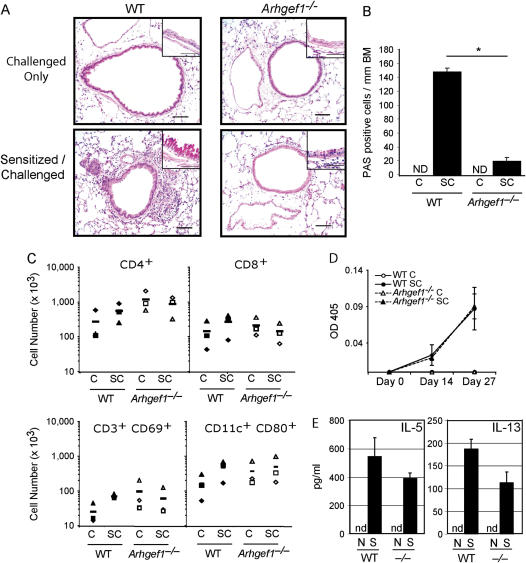
Figure 2.
Sensitized Arhgef1−/− mice mount a normal systemic immune response, but display only minor infiltration in lung after airway challenge. (A) Hematoxylin and eosin–stained lung sections from sensitized and challenged wild-type (WT) and mutant mice. Original magnification of insets: ×100 of periodic acid Schiff (PAS)–stained sections. Bars = 50 μm. (B) Number of PAS-positive cells/mm of basement membrane (BM) were identified from paraffin sections of sensitized and challenged lungs. ND = none detected. *p < 0.05 (C) Number of WT and Arhgef1−/− cells in indicated lung leukocyte populations after sensitization and/or challenge. Leukocytes were isolated from enzymatic lung digests and individual populations identified by flow cytometry, as described in Methods. Filled symbols are WT and open symbols are Arhgef1−/−. CD4 and CD8 T cells identified as CD3+CD4+ or CD3+CD8+, respectively. Symbols represent individual experiments and are the average number from three mice per experiment. (D) OVA-specific IgE levels were determined in serum collected on Days 0, 14, and 27 of the OVA protocol from WT and Arhgef1−/− mice. Open symbols represent challenged-only mice and filled symbols represent sensitized and challenged mice. (E) After sensitization, levels of IL-5 and IL-13 were quantified from supernatants taken from splenocyte cultures. N = naive; S = sensitized; nd = not detected.

