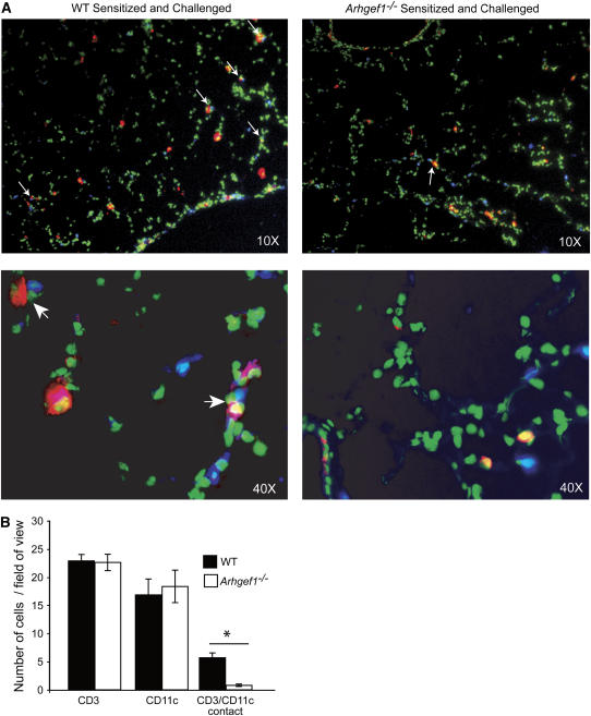
Figure 4.
Reduced CD3+ and CD11c+ cell interaction in lung tissue of sensitized and challenged Arhgef1−/− mice. (A) Histologic lung sections stained with anti-CD3 (blue) and anti-CD11c (red). Green color is autofluorescence and is used to identify lung tissue. Top panels are ×10 original magnification and bottom panels ×40 original magnification. Arrows indicate CD3+/CD11c+ contacts. (B) Number of CD3+ T cells, CD11c+ cells, and CD3+-CD11c+ cell contacts per 10× field of view in sensitized and challenged wild-type (WT) and Arhgef1−/− mice. Average numbers were obtained from at least four fields of view per section from four different sections per set of lungs and from four individual mice. *p < 0.05 by Student's t test.

