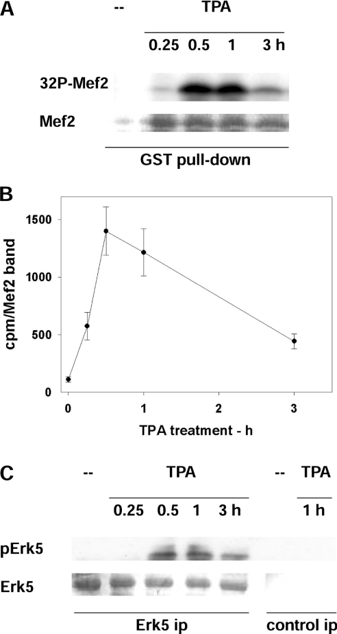
Figure 5. Activation of ERK5 in cells treated with PMA (TPA).
(A, B) ERK5 activity assayed as Mef2 kinase. (A) GST–Mef2 was bound to glutathione–Sepharose beads and incubated with [γ-32P]ATP/MgCl2 and cell extract from rat fibroblasts exposed to 300 nM PMA for the indicated time. This was followed by electrophoresis, Coomassie Blue staining (Mef2) and exposure to detect the radioactivity incorporated into Mef2 (32P-Mef2). (B) Quantification of 32P-Mef2. Mef2 bands were excised from the gel (see A) and counted (c.p.m./Mef2 band). Results are means±S.E.M. for three independent experiments. (C) ERK5 activity assayed as pERK5. ERK5 was immunoprecipitated (ERK5 ip), followed by electrophoresis, probing with antibodies that recognize the phosphorylation of Thr218 and Tyr220 of ERK5 (pERK5) and membrane staining with Coomassie Blue (ERK5). Control immunoprecipitates (control ip) are also shown.

