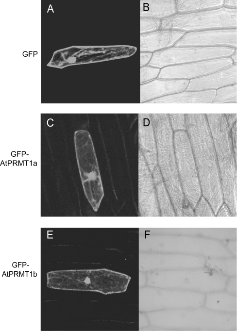
Figure 4. AtPRMT1a and AtPRMT1b localize to the nucleus as well as the cytoplasm.
Both AtPRMT1a and AtPRMT1b were fused to the C-terminal end of GFP and transiently transfected into onion epidermal cells. Expression of GFP (A, B), GFP–AtPRMT1a (C, D) and GFP–AtPRMT1b (E, F) were observed by confocal fluorescence microscopy (A, C and E) and brightfield microscopy (B, D and F).

