
Fig 5.
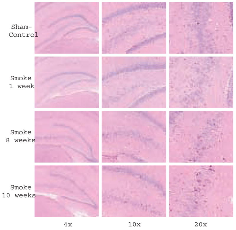
Delayed neuronal injury after inhalation of smoke. Histochemical analysis of coronal sections from smoke and sham-smoke treated rats: Representative photomicrographs of hematoxylin and eosin staining through the hippocampus (Bregma -3.14) captured with 4x, 10x and 20x objectives, from control rats and rats sacrificed 1, 8 and 10 weeks post smoke exposure. An increase in number of dark neurons (dark pink) is observed at 8 and 10 weeks.