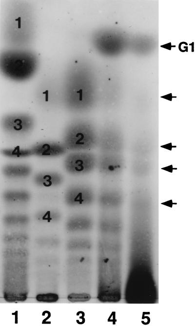
Figure 3.
TLC analysis of the products of mild acid hydrolysis of the receptor. Approximately 40 μg as Sia was spotted on a silica gel plate, developed with 1-propanol/25% ammonia/water (6:1:2.5) for 12 h, and visualized by the resorcinol reagent (22). The following polySia-related molecules were hydrolyzed under the conditions indicated in parenthesis. Lanes: 1, colominic acid (pH 4.5, 80°C, 3 h); 2, rainbow trout polysialoglycoprotein (pH 4.5, 80°C, 3 h); 3, sea urchin S. purpuratus polySia-gp (0.1 M TFA, 65°C, 20 min); 4, H. pulcherrimus ESP-Sia (pH 4.5, 80°C, 5 h); 5, 350-kDa receptor protein (pH 4.5, 80°C, 15 h). The dark spot at the origin is not a resorcinol positive substance since it is brown, rather than purple. Numbers in lanes 1-3 represent the degree of polymerization. G1 in lane 5 is (SO4−)-9Neu5Gc. The arrowheads indicate the eluted position of NeuGc monomer (Upper) and α2→5-Oglycolyl-linked Neu5Gc di, tri, and tetramer (Lower).

