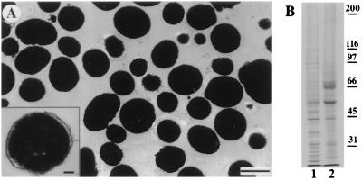
Figure 1.
(A) Electron micrograph of a thin-sectioned pellet of purified melanosomes. The purified melanosome fractions are uncontaminated by other organelles. (Bar = 0.5 μm.) (Inset) A melanosome at higher magnification showing bilayer membrane encapsulating the electron-dense melanin core. (Bar = 0.1 μm.) (B) A Coomassie-stained polyacrylamide gel comparing melanophore extract (lane 1) with purified melanosomes (lane 2). The positions of the molecular weight markers are indicated to the right of lane 2.

